Multimodal MRI assessment for first episode psychosis- A major change in the thalamus and an efficient stratification of a subgroup
Abstract
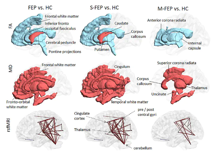
Multi-institutional brain imaging studies have emerged to resolve conflicting results among individual studies. However, adjusting multiple variables at the technical and cohort levels is challenging. Therefore, it is important to explore approaches that provide meaningful results from relatively small samples at institutional levels. We studied 87 first episode psychosis (FEP) patients and 62 healthy subjects by combining supervised integrated factor analysis (SIFA) with a novel pipeline for automated structure-based analysis, an efficient and comprehensive method for dimensional data reduction that our group recently established. We integrated multiple MRI features (volume, DTI indices, resting state fMRI-rsfMRI) in the whole brain of each participant in an unbiased manner. The automated structure-based analysis showed widespread DTI abnormalities in FEP and rs-fMRI differences between FEP and healthy subjects mostly centered in thalamus. The combination of multiple modalities with SIFA was more efficient than the use of single modalities to stratify a subgroup of FEP (individuals with schizophrenia or schizoaffective disorder) that had more robust deficits from the overall FEP group. The information from multiple MRI modalities and analytical methods highlighted the thalamus as significantly abnormal in FEP. This study serves as a proof-of-concept for the potential of this methodology to reveal disease underpins and to stratify populations into more homogeneous sub-groups.
Chenfei Ye
Research Associate Professor
The brain’s billions of neurons resemble trees of many species and come in many fantastic shapes. Only the most determined explorers can hope to capture a glimpse of this forest’s interior, and even they see little, and see it poorly.